Wednesday 15 August 2012

Case 3 - Infected NonUnion Radius Ulna Forearm


A 42 year old homemaker, mother of 2, sustained a closed fracture of the radius ulna in February 2011.

She saw an orthopedic surgeon close to her home, and an open reduction and internal fixation by plates was done  on the same day.

Immediate Postoperative Xray
On the second day after surgery, she developed a high fever. Blood counts were asked for by the first surgeon, and a WBC count of 13500 was noted.
The Antibiotic was changed - presumably, a higher antibiotic was administered.
This settled the fever somewhat, but the woulnd was noted to be 'gaping'. This was secondarily sutured on the 5th day and she was sent home.

However, the patient continued to be 'ill' and had pain & swelling in the operated limb, and decided to take a second opinion.
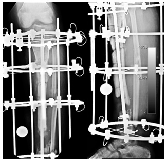
The second surgeon removed the radial plate and put on an external fixator on the radius.
 The ulnar plate was retained.

Radial plate removed and Fixator applied
She was administered oral antibiotics.
She continued to be troubled by discharge from the radial incision and another 3 weeks later the ulnar incision began discharging seropurulent fluid.
This was tackled by removal of the ulnar plate and application of a fixator.

Ulnar plate removed and fixator applied
Unfortunately - this didn't solve the problem either.

She was asked to get a scan done, and when she came to the scan center which is in the same building she apparently saw the CLLR board which specifically lists infection/osteomyelitis as one of our areas of expertise, and has a line "Cutting edge techniques tempered by 25 years of experience".
That prompted her to seek our opinion.

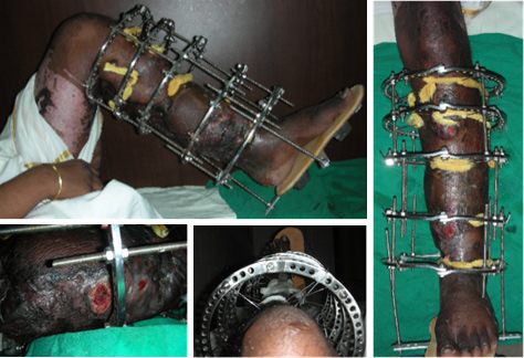
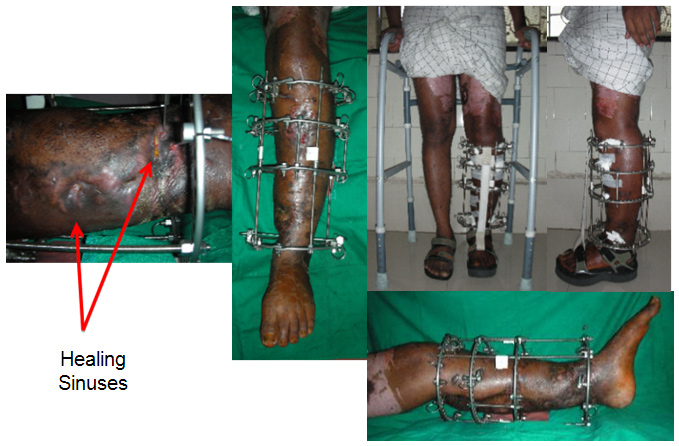
Pinsites get worse, Radius sequestrating

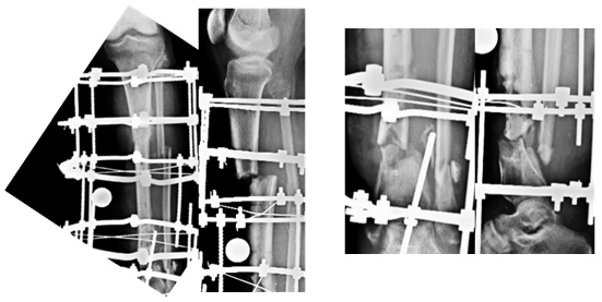
X-ray at presentation
She had no other co-morbid conditions that could explain the florid infection.
After reviewing her history, reviewing her xrays and examining her, we sat her down (along with husband and a couple of relatives) and discussed the options before her.

Clinical Appearance and pinsites at presentation
Her Xrays showed that the middle of the radius had practically separated, and was surely a sequestrum, with probably similar changes in the ulna too.
She seemed to have a 'wrist drop' which I thought was more because of adhesions or the inflamation within the compartment, rather than a problem with the radial or posterior interosseous nerve.
The only option really in my opinion was to do a proper debridement, clear the infection and then think of the reconstructive aspect.

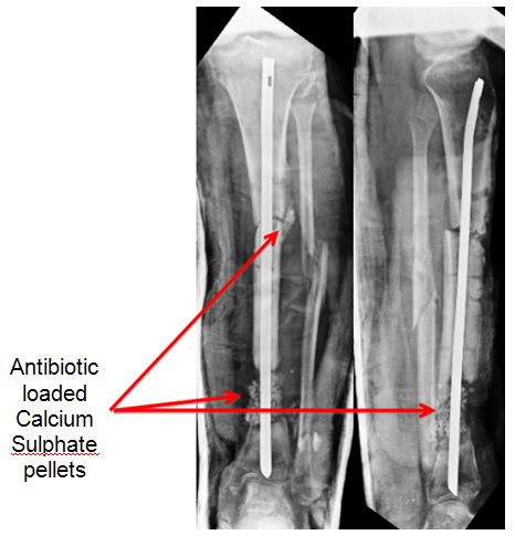
In May 2011, we did the surgery for her and the plan was to do a thorough debridement, use local antibiotic in Calcium Sulphate pellets (Stimulan), and stabilise.
The debridement and the antibiotic pellets was the 'easy' part. How to stabilise a large defect (for I was pretty sure that was what we were going to have at the end of debridement.

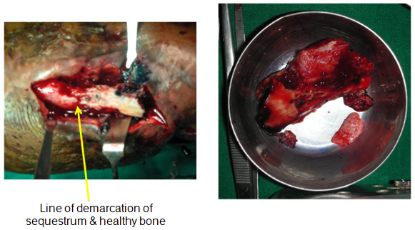
Central diaphysis of the Radius and Ulna sequestrated
Once the necrotic bone, granulation etc was removed, we decided to use TENS nail, along with external support by a plaster slab - basically because in case we needed to use a VAC in the postop period, a fixator would have made it difficult to apply the VAC.
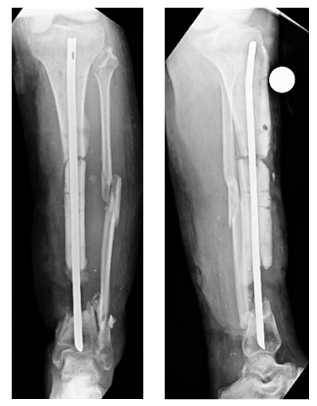
Also, having had a fair amount of experience with the TENS nail, I was certain of a reasonable stability as long as the right size and right length (to seat it well upto subchondral bone) was used.

We put in the nails and took care to seat the ends as close to the subchondral bone as we could, thereby gaining some rotational stability. At the lower end of the radius, I buried the end into the bone to provide some resistance against collapse.

Surprisingly (maybe not so surprisingly), with the nails in the forearm was quite stable, to rotation and also preventing collapse of the gap.
We filled in the empty space with Stimulan loaded with 2 gms vancomycin and 3 million units Colistin (based on the earlier culture reports) and sent off tissue samples from multiple areas in the wound for culture (which would decide our IV antibiotics.

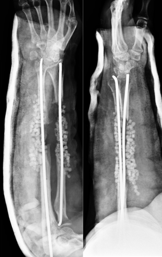
Stabiised by TENS nail. Stimulan Pellets for local Antibiotic Delivery
Postoperatively, the forearm quite clearly 'calmed down'. Regular dressings showed no problems, and blood parameters improved gradually.

Incisions 2 days and 7 days after surgery
She was on IV antibiotics based on the last culture that was sent from the deep tissues.

I have been minimising the use of IV antibiotics based on a combination of clinical appearance and response, and blood parameters - notably CRP. Based on these, we stopped IV antibiotics after 3 weeks, despite the CRP not being normal, but it was showing a clear downward trend and the patient was comfortable.
 No further antibiotics were administered to her after this. 

Healing of the Incisions
Over the next few months, we kept a regular watch on her, clinically as well as by xray.

The TENS nails did their job of stabilisation well, and xrays showed gradual disappearance of the pellets (as expected).

Pellets Dissapear over time. TENS nail holds position well
We had discussed with the patient, and suggested that a Vascular Fibula was the best option for her to cut short the recovery and maintain function (in contrast to the Ilizarov, which would have been the other option)

Our plastic surgeons were happy to do a double barreled fibula, and we fixed it with a long titanium plate that almost covered the bones completely.

Vascularised Fibula with Pedicle, Plate fixation thru single incision

Vascularised Fibula with Skin paddle
 For about 6 weeks she was protected in a slab, with intermittent mobilisation out of the slab, and gradual weaning off of the external support after that.

Double Barrelled Fibula and LCP fixation
Today at 1 year after that surgery, she is back to an active life, happy (though she admits that entering any hospital still makes her a little nervous and uncomfortable), fairly good function from a medical standpoint, but "able to do everything that she needs to do" and the xrays show a solid union at all the four sites, without any additional procedures being required.

Excellent Function, except for pronation beyond midprone
Solidly Healed on Xray
This particular patient, really underlines the importance of
1. the recognition of the destructiveness of infection,
2. of the need for experience to deal with it aggressively, 
3. of the requirement of the surgeon to deal with the surgery, the pharmacology and the psychology of infection.
4. the need to use the best knowledge, techniques, and tools/implants available to achieve an optimal result.

Sunday 20 May 2012

Case 2 - Forearm Deformity

Clinical Picture and movements at presentation

This 15 year old girl was a precious child for her parents, born after many attempts of IVF. 

She had a short and bowed forearm, and in addition had an unsightly prominence in the lateral aspect of the elbow. 

She also complained of pain when she had to do any writing tasks for a long time. 

Xray at presentation

Xrays revealed an exostosis at the distal ulna, which caused a short ulna, which in turn by the check rein effect caused an exaggerated bowing of the radius and dislocation of the head of the radius. Some surgical procedure had been attempted in the past, details of which were unavailable.

The challenge here was to correct the bowing deformity, as well as the prominence of the radial head, without much surgical scars.

We first excised the ulnar exostosis. 6 weeks after that, we applied a pediatric LRS fixator on the ulna.
The trick here was to put in the proximal and distal set of pins without the rail, such that each set of pins was perpendicular to that segment of bone.

C arm pictures of fixator application & osteotomy

Once the clamps were fixed to the rails, the bowing of the ulna was automatically corrected.

Due to the acute correction, instead of the normal 7 days, we began lengthening after 10 days, and continued lengthening, till the distal end of the ulna reached the carpus / distal end of radius.

Sequential Xrays of Ulnar Lengthening
Once the Ulna was at the desired length, she was taken to theatre, and the distal end of the ulna was locked or coupled to the distal end of the radius, by means of 1 screw and 1 K wire, put in percutaneously.

C arm pictures of insertion of Screw and K wire
The ulnar lengthening was then continued, till the radial head prominence disappeared. No attempt was made to reduce the radial head, because even preoperatively, with the gross dislocation of the radial head, she had a good range of movements. We anticipated that she would retain good movement, and IF required, an excision of the radial head could be done later.

The Radial Head moves distally
Function was encouraged while in the fixator, and when the regenerate appeared healed well, the fixator was removed.

Function with the Fixator
Healed regenerate
She had good function equal to her preoperative status even at 1 week post removal
Scars and Prono-Supination
The bone was healed well as confirmed on various views.


At her one year followup, she is obviously a happy patient, free of her deformity, and her complaints.
Notice the the bump of the radial head is hardly (if at all) visible.


She is now back at her village, attending Junior College.

Sunday 13 May 2012

Case 1 - Continued

Case 1 continued . .

Corticotomy

Once we were sure that the infection was settled, (and the circumference of the leg reduced significantly), we went ahead with adding an ilizarov fixator to the limb, and doing a corticotomy in the proximal tibia. 


There were two nonunions we were dealing with, one proximal in the midshaft, and another gap, distally. 

The frame was constructed to allow continued compression across the proximal nonunion site (if you look closely, you will see the arced wires in the xrays and clinical pictures). 

The corticotomy posed a challenge, with the nail already in, and the limb not really a good one to do a Gigli Saw osteotomy on. 

After the a period of 10 days, distraction (transport) was begun. As the osteotomy site opened up, the gap at the lower nonunion closed. 

Improving the Docking site

At one point, the two edges of the gap nonunion came into contact, and would have prevented a good cross section of bone for the healing. Therefore we did a percutaneous osteotomy and osteotomised, in situ the long beak of the proximal fragment such that it could now present a broader surface for the docking.


After docking was achieved, proximal lengthening was continued for a couple of centimetres to equalise his length. 


The Gap nonunion healed without any bone grafting, the regenerate healed without any issues, and the patient has good function. No discharge. 


I am told he will be getting married soon, and we will certainly celebrate with him. 


TEAM CLLR

Case 1 - Infected Nonunion Tibia

Case 1 - Infected Nonunion Tibia


At Presentation
25 year old young man, had a bad accident about 10 months back. The tibia was fractured in two places (segmental fracture) with injury to the blood vessel and nerve. He underwent a number of surgeries namely arterial grafting, muscle flap, skin grafting, Ilizarov fixation before presenting to us with discharging sinuses, inability to walk and lack of confidence.


X-rays at Presentation
The x-rays showed nonunion at both the upper and lower fracture sites. The lower fracture site showed infection and dead bone. The fixator was not holding the bone fragments in good alignment and the wires were all loose and some of them were infected.


PET Scan
The FDG PET Scan showing active infection at the lower fracture site. An arterial DSA (Digital Subtraction Angiogram) was done to determine the course and patency of the arteries.


Sequestrum – Dead Bone
The dead bone was excised. The line of demarcation between the dead and viable bone is shown here in the picture on the left (yellow arrow).


In surgery the dead bone (sequestrum) & all the infective tissue was removed. An intra-medullary V-nail was applied to keep the bone aligned and antibiotic loaded calcium sulphate pellets were instilled in the proximal and distal fracture sites to control infection.


4 Weeks
The calcium sulphate pellets are completely absorbed. The infection is settled and the sinuses healing well. The upper fracture is showing signs of healing.


Bone Transport
The Ilizarov fixator is applied while the intra-medullary nail is still inside. An osteotomy was done in the proximal aspect for the purpose of bone transport.


5 Weeks